Broken bone in foot healing. Broken Foot Healing: Expert Guide to Diagnosis, Treatment, and Recovery
How long does it take for a broken foot to heal. What are the symptoms of a broken foot. How is a broken foot diagnosed and treated. What does the recovery process for a broken foot involve. How can you prevent foot fractures.
Understanding Broken Foot Injuries: Types and Causes
Foot fractures can range from minor cracks to severe breaks that completely sever the bone. In some cases, the broken bone may even pierce the skin, resulting in what’s known as an open fracture. Understanding the various types of foot fractures is crucial for proper diagnosis and treatment.
Common Types of Foot Fractures
- Stress fractures: Tiny cracks in the bone caused by repetitive force or overuse
- Avulsion fractures: When a small piece of bone is pulled away by a tendon or ligament
- Comminuted fractures: The bone breaks into multiple pieces
- Displaced fractures: The broken ends of the bone are separated and misaligned
- Non-displaced fractures: The bone cracks but maintains its proper alignment
Foot fractures can occur due to various causes, including high-impact accidents, falls, sports injuries, or even repetitive stress over time. Certain medical conditions, such as osteoporosis, can also increase the risk of foot fractures.
Recognizing the Symptoms of a Broken Foot
Identifying a broken foot can be challenging, especially if there’s no visible deformity or open wound. However, several key symptoms can indicate a potential fracture:
- Sudden, intense pain at the site of injury
- Swelling and bruising
- Difficulty walking or bearing weight on the affected foot
- Visible deformity or misalignment of the foot or toes
- A grinding or snapping sound at the time of injury
- Tenderness or pain when touching the injured area
- Numbness or tingling sensations
Can you have a broken foot without realizing it? In some cases, minor fractures or stress fractures may not cause severe pain immediately, leading to delayed diagnosis. It’s essential to pay attention to persistent foot pain or discomfort, even if there’s no apparent injury.
When to Seek Medical Attention for a Suspected Foot Fracture
While minor toe fractures can often be treated at home, it’s crucial to seek immediate medical attention for suspected fractures in the foot or big toe. Certain symptoms warrant urgent care:

- Visible deformity or misalignment of the foot or toes
- Open wounds or broken skin near the injury site
- Cold, numb, or tingling sensations in the toes or foot
- Bluish or grey discoloration of the toes or foot
- A crushed or severely injured foot
- Inability to walk or bear weight on the affected foot
- Persistent pain or swelling that doesn’t improve with home treatment
Is it possible to differentiate between a sprain and a fracture? While both conditions can cause similar symptoms, fractures typically result in more intense and continuous pain. Additionally, swelling and bruising tend to be more severe in fractures compared to sprains. However, only a medical professional can provide an accurate diagnosis through imaging tests.
Diagnosis and Imaging Techniques for Foot Fractures
Proper diagnosis of a foot fracture is crucial for determining the appropriate treatment plan. Healthcare providers employ various diagnostic techniques to assess the extent and location of the injury:
Common Diagnostic Methods
- Physical examination: The doctor will inspect the foot for visible signs of injury and assess pain levels and range of motion.
- X-rays: These are the most common imaging tests used to diagnose foot fractures, providing clear images of bone structures.
- CT scans: For more complex fractures, CT scans offer detailed 3D images of the bones and surrounding tissues.
- MRI scans: These can help detect stress fractures or soft tissue injuries that may not be visible on X-rays.
- Bone scans: In some cases, a bone scan may be used to identify stress fractures or other subtle bone abnormalities.
How accurate are X-rays in diagnosing foot fractures? While X-rays are generally reliable for identifying most fractures, some small or stress fractures may not be immediately visible. In such cases, additional imaging tests or follow-up X-rays may be necessary for a definitive diagnosis.

Treatment Options and Recovery Process for Broken Feet
The treatment approach for a broken foot depends on the location and severity of the fracture. In most cases, the primary goal is to immobilize the affected area and promote proper bone healing:
Conservative Treatment Methods
- Rest and limited weight-bearing
- Ice therapy to reduce swelling and pain
- Compression bandages to minimize swelling
- Elevation of the affected foot
- Pain management with over-the-counter or prescribed medications
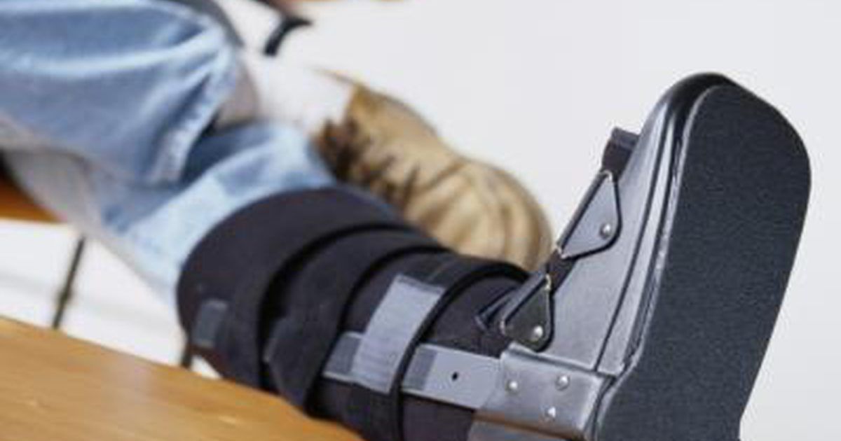
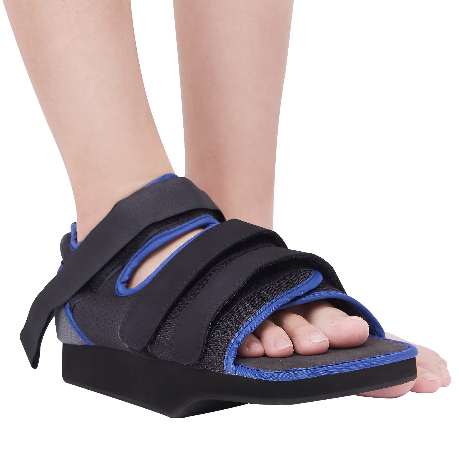
- Immobilization using casts, splints, or special footwear
- Crutches or other mobility aids to avoid putting weight on the injured foot
Surgical Interventions
In some cases, particularly for severe or complex fractures, surgery may be necessary. Surgical options may include:
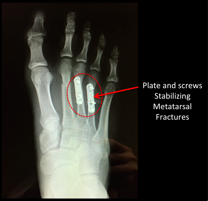
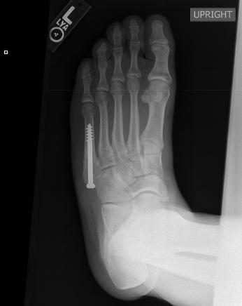
- Internal fixation: Using screws, plates, or rods to hold bone fragments in place
- External fixation: Applying an external frame to stabilize the bones
- Bone grafting: Adding bone tissue to promote healing in complex fractures
What factors influence the choice between conservative treatment and surgery? The decision depends on various factors, including the type and location of the fracture, the patient’s overall health, and their activity level. Your healthcare provider will recommend the most appropriate treatment based on your individual case.
Healing Time and Rehabilitation for Foot Fractures
The healing process for a broken foot can vary depending on the severity of the fracture and the individual’s overall health. On average, most foot fractures take about 4-6 weeks to heal, but some may require up to 10-12 weeks for complete recovery.
Factors Affecting Healing Time
- Age and overall health of the patient
- Severity and location of the fracture
- Adherence to treatment plans and follow-up care
- Nutrition and lifestyle factors
- Presence of underlying medical conditions
How can you promote faster healing of a broken foot? While you can’t rush the natural healing process, you can support it by following your doctor’s instructions, maintaining a healthy diet rich in calcium and vitamin D, avoiding smoking, and gradually increasing weight-bearing activities as advised by your healthcare provider.
Rehabilitation and Physical Therapy
As the fracture heals, your doctor may recommend physical therapy to help restore strength, flexibility, and function to your foot. Rehabilitation may include:
- Range of motion exercises
- Strength training for foot and ankle muscles
- Balance and proprioception exercises
- Gait training to improve walking patterns
- Gradual return to normal activities and sports
When is it safe to return to normal activities after a foot fracture? The timeline for returning to regular activities varies depending on the individual and the nature of the fracture. It’s crucial to follow your doctor’s guidance and avoid rushing the recovery process to prevent re-injury or complications.
Preventing Foot Fractures: Tips and Best Practices
While not all foot fractures can be prevented, there are several steps you can take to reduce your risk of injury:
Lifestyle and Safety Measures
- Wear proper footwear that provides adequate support and protection
- Use appropriate safety equipment during sports and high-risk activities
- Maintain a healthy diet rich in calcium and vitamin D to support bone health
- Engage in regular weight-bearing exercises to strengthen bones
- Be cautious when walking on uneven or slippery surfaces
- Gradually increase the intensity and duration of physical activities
- Address any underlying medical conditions that may affect bone health
How effective are these preventive measures in reducing the risk of foot fractures? While no method can guarantee complete prevention, consistently following these guidelines can significantly lower your risk of experiencing a foot fracture. It’s especially important for individuals with a higher risk of fractures, such as those with osteoporosis or a history of previous fractures, to prioritize these preventive measures.
Long-Term Outlook and Potential Complications of Foot Fractures
Most foot fractures heal successfully with proper treatment and care. However, it’s essential to be aware of potential long-term effects and complications that may arise:
Possible Long-Term Effects
- Chronic pain or stiffness in the affected area
- Arthritis in the injured joint
- Altered gait or walking pattern
- Increased risk of future fractures in the same area
- Nerve damage or numbness
Potential Complications
- Nonunion: When the broken bone fails to heal properly
- Malunion: When the bone heals in an incorrect position
- Infection, particularly in cases of open fractures
- Complex regional pain syndrome (CRPS)
- Blood clots or deep vein thrombosis due to immobilization
Can foot fractures lead to permanent disability? While most foot fractures heal without long-term consequences, severe or complex fractures may sometimes result in permanent changes in foot function or mobility. Proper treatment, rehabilitation, and follow-up care are crucial to minimize the risk of long-term complications.

Understanding the diagnosis, treatment, and recovery process for foot fractures is essential for anyone who experiences this type of injury. By recognizing the symptoms, seeking timely medical attention, and following proper treatment protocols, most individuals can expect a full recovery and return to normal activities. Additionally, implementing preventive measures can help reduce the risk of future foot fractures and maintain overall foot health.
Symptoms, what does it look like, recovery, and treatment
Broken foot treatment may involve rest, applying ice, elevating the foot, taking anti-inflammatory medications, wearing a cast, or using crutches to keep weight off of the bones until they heal.
Healing times for a broken foot are often around 4–6 weeks, but may take longer in some cases.
This article looks at the causes and symptoms of a broken foot and when to seek medical help. It also discusses first aid, diagnosis, treatment, recovery, and prevention tips.
Bones can break in many ways. These can vary from small cracks and splinters to complete breaks that sever the bone. Severe breaks can tear or pierce the skin and leave a wound. These are known as open fractures.
If there is no visible displacement of the bone or a clear wound, a person may not be able to tell if a bone has broken. Also, some minor cracks or breaks may not result in much pain.
Deformity of a toe or an area of the foot, such as an unusual bulge, strongly indicates a break.
Other indications of a broken bone in the foot include:
- hearing or feeling a snap or grinding noise when an injury happens
- pain or difficulty moving the foot
- pain or trouble walking or bearing weight on the foot
- tenderness or pain when touching the injury
- feeling faint, dizzy, or sick following the injury
What does a fractured foot look like?
When to contact a doctor
A person should seek immediate medical assistance if they suspect they have broken a bone in their foot or big toe. They should not attempt to drive. Broken smaller toes are less severe, and a person should attempt to treat them at home first.
A person should also seek immediate assistance if:
- the leg, foot, or toe is deformed or pointing the wrong way
- there is a wound or broken skin near the injury
- the toes or foot are cold, numb, or tingling
- the toes or foot have turned blue or grey
- the foot is crushed
A person should also contact a doctor for any injury that prevents walking or causes persistent pain or swelling in the feet.
Injuries to the foot may cause a sprain rather than a break. A sprain is when the tissue between the bones, known as ligaments, tear, or stretch.
Sprains can result in similar symptoms to breaks, and it may be difficult for a person to tell the two conditions apart.
In general, a broken foot will result in more intense and continuous pain. In addition, swelling and bruising in the foot will typically be more severe if it is due to a break rather than a sprain.
However, sprains can still cause distress and impair a person’s movement. A doctor will order imaging tests to accurately diagnose whether a person’s injury is a break or a sprain.
Learn more about the differences between sprains and strains here.
A broken foot or toe may take 4–6 weeks to heal fully. However, in some cases, healing time can be as long as 10–12 weeks.
Recovering individuals should follow the RICE method and any specific instructions from their doctor. A person may also require follow-up X-rays or other scans to ensure proper healing and alignment.
Returning to physical activity too soon can risk poor healing, re-injury, or a complete fracture. A person should contact a doctor if the pain or swelling returns.
People should always seek medical attention if they suspect they have broken a bone in their foot or big toe.
However, immediately following an injury, they may benefit from following the RICE principle while seeking or waiting for help. The acronym stands for:
- Rest: A person who suspects they have broken a bone should keep pressure off the injured foot or limit weight bearing until it gets better, or a doctor can examine it. Unnecessary walking could worsen the injury.
- Ice: Immediately apply ice to the injury to reduce pain and swelling. A person may use icepacks for up to 20 minutes several times a day for the first 48 hours. They should not apply ice packs directly to the skin.
- Compression: A person should wrap the foot in a soft dressing or bandage.
 However, they must ensure the bandage is not too tight, as this may stop the blood from circulating.
 - Elevation: Elevate the foot, as much as possible, with pillows. Ideally, a person will raise the foot above the level of the heart. This also helps with pain and swelling.
Following breaks in smaller toes, a person can tape a broken toe to an adjacent, uninjured toe for support. Some people refer to this as ‘buddy taping.’ This involves placing a piece of cotton wool or gauze between the two toes, then securing them together with surgical tape.
A person may be able to relieve immediate pains by taking over-the-counter pain relief medication, such as acetaminophen or ibuprofen. If walking on a broken foot or toe becomes necessary, the individual should wear a wide, sturdy shoe that does not pressure the injured area.
A person can use the RICE principle to treat a strain or sprain in the foot or ankle.
Although the foot can typically withstand considerable force, breaking bones in the foot or toes is common.
A broken foot can result from simply stumbling, tripping, or kicking something. Twisting the foot or ankle awkwardly by falling or being hit by a heavy object can also break a bone.
Stress fractures are a particular risk in athletes or anyone who partakes in high-impact sports, such as football, basketball, running, or dancing.
These are tiny, sometimes microscopic, cracks that can enlarge over time. They tend to be caused by repetitive activities or sudden increases in exercise intensity.
To diagnose a broken foot, a doctor will ask questions about the injury and feel and manipulate the affected foot. They may order an X-ray to confirm or further assess a possible break.
A suspected stress fracture may require an MRI or ultrasound, as these tiny fractures can be difficult to detect on an X-ray.
To reduce the risk of injuring the feet, people should keep the floors at home and in the workplace free of clutter. Those working on construction sites or in other hazardous environments should wear professional safety boots.
When partaking in sports or exercise, the following advice can help prevent stress fractures and other foot injuries:
- use shoes and equipment appropriate to the activity
- stretch, warm up, and start the activity slowly
- gradually increase speed, time, distance, or intensity of a new activity or after a break
- use stretches and exercises to build up the calf muscles
- alternate with low-impact activities, such as swimming and cycling
- eat foods rich in calcium and vitamin D to build up bone strength
The symptoms of a broken foot are often similar to those of a strain or sprain. However, swelling, pain, and visual deformity will typically be greater following a bone break.
A person may break a bone in their foot through an impact injury or overuse. Breaks can vary from small cracks or splinters to open fractures where a portion of bone breaks through the skin.
Anyone who suspects they have broken a bone in their foot should contact a doctor immediately.
A person may be able to treat breaks in smaller toes at home with buddy taping and the RICE principle.
Below are answers to some frequently asked questions about a broken foot.
What happens if you don’t treat a broken foot?
Bones may heal out of natural alignment if a person does not seek medical treatment. This can lead to permanent bone deformity and mobility problems. If a person has an open fracture and does not seek treatment, they may also be at risk of developing an infection in the wound.
Does a broken foot always bruise?
A person may break a bone within their foot without experiencing any visible bruising. Bruises are the result of blood pooling under the skin. Overuse or injury can lead a bone to break without disturbing blood vessels.
Symptoms, what does it look like, recovery, and treatment
Broken foot treatment may involve rest, applying ice, elevating the foot, taking anti-inflammatory medications, wearing a cast, or using crutches to keep weight off of the bones until they heal.
Healing times for a broken foot are often around 4–6 weeks, but may take longer in some cases.
This article looks at the causes and symptoms of a broken foot and when to seek medical help. It also discusses first aid, diagnosis, treatment, recovery, and prevention tips.
Bones can break in many ways. These can vary from small cracks and splinters to complete breaks that sever the bone. Severe breaks can tear or pierce the skin and leave a wound. These are known as open fractures.
If there is no visible displacement of the bone or a clear wound, a person may not be able to tell if a bone has broken. Also, some minor cracks or breaks may not result in much pain.
Deformity of a toe or an area of the foot, such as an unusual bulge, strongly indicates a break.
Other indications of a broken bone in the foot include:
- hearing or feeling a snap or grinding noise when an injury happens
- pain or difficulty moving the foot
- pain or trouble walking or bearing weight on the foot
- tenderness or pain when touching the injury
- feeling faint, dizzy, or sick following the injury
What does a fractured foot look like?
When to contact a doctor
A person should seek immediate medical assistance if they suspect they have broken a bone in their foot or big toe. They should not attempt to drive. Broken smaller toes are less severe, and a person should attempt to treat them at home first.
A person should also seek immediate assistance if:
- the leg, foot, or toe is deformed or pointing the wrong way
- there is a wound or broken skin near the injury
- the toes or foot are cold, numb, or tingling
- the toes or foot have turned blue or grey
- the foot is crushed
A person should also contact a doctor for any injury that prevents walking or causes persistent pain or swelling in the feet.
Injuries to the foot may cause a sprain rather than a break. A sprain is when the tissue between the bones, known as ligaments, tear, or stretch.
Sprains can result in similar symptoms to breaks, and it may be difficult for a person to tell the two conditions apart.
In general, a broken foot will result in more intense and continuous pain. In addition, swelling and bruising in the foot will typically be more severe if it is due to a break rather than a sprain.
However, sprains can still cause distress and impair a person’s movement. A doctor will order imaging tests to accurately diagnose whether a person’s injury is a break or a sprain.
Learn more about the differences between sprains and strains here.
A broken foot or toe may take 4–6 weeks to heal fully. However, in some cases, healing time can be as long as 10–12 weeks.
Recovering individuals should follow the RICE method and any specific instructions from their doctor. A person may also require follow-up X-rays or other scans to ensure proper healing and alignment.
Returning to physical activity too soon can risk poor healing, re-injury, or a complete fracture. A person should contact a doctor if the pain or swelling returns.
People should always seek medical attention if they suspect they have broken a bone in their foot or big toe.
However, immediately following an injury, they may benefit from following the RICE principle while seeking or waiting for help.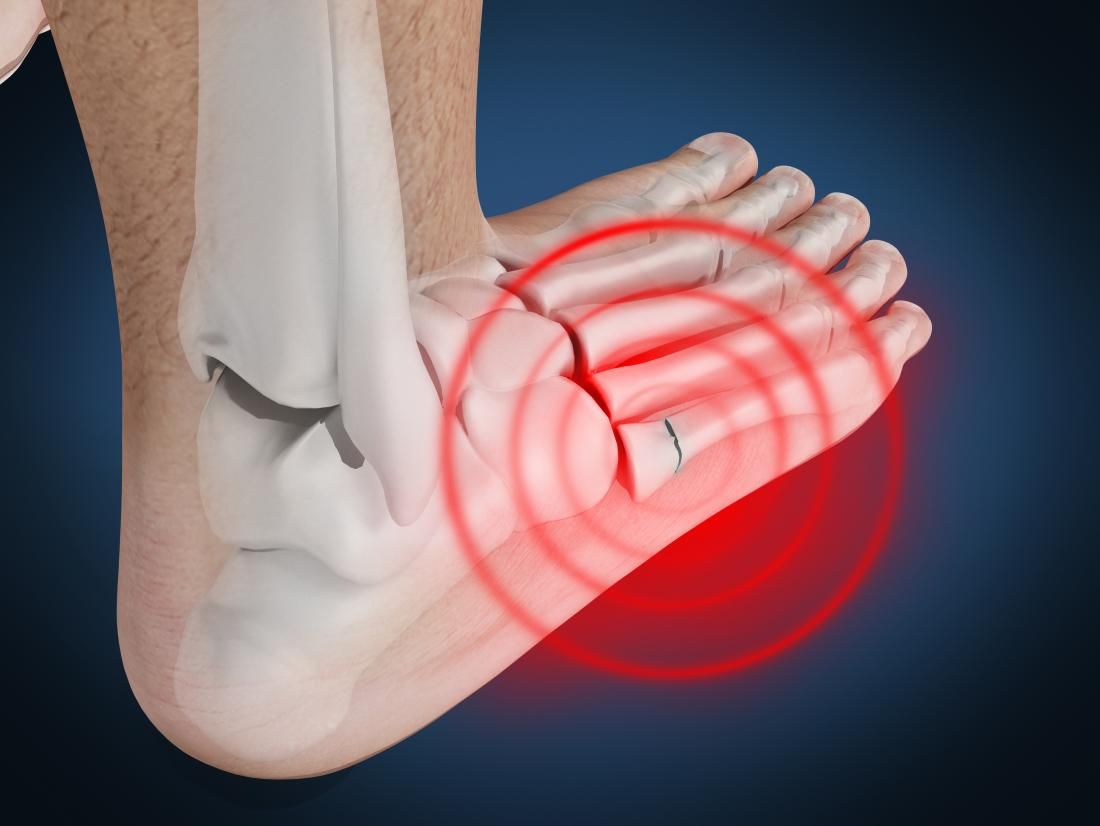 The acronym stands for:
- Rest: A person who suspects they have broken a bone should keep pressure off the injured foot or limit weight bearing until it gets better, or a doctor can examine it. Unnecessary walking could worsen the injury.
- Ice: Immediately apply ice to the injury to reduce pain and swelling. A person may use icepacks for up to 20 minutes several times a day for the first 48 hours. They should not apply ice packs directly to the skin.
- Compression: A person should wrap the foot in a soft dressing or bandage. However, they must ensure the bandage is not too tight, as this may stop the blood from circulating.
- Elevation: Elevate the foot, as much as possible, with pillows. Ideally, a person will raise the foot above the level of the heart. This also helps with pain and swelling.
Following breaks in smaller toes, a person can tape a broken toe to an adjacent, uninjured toe for support. Some people refer to this as ‘buddy taping.’ This involves placing a piece of cotton wool or gauze between the two toes, then securing them together with surgical tape.
A person may be able to relieve immediate pains by taking over-the-counter pain relief medication, such as acetaminophen or ibuprofen. If walking on a broken foot or toe becomes necessary, the individual should wear a wide, sturdy shoe that does not pressure the injured area.
A person can use the RICE principle to treat a strain or sprain in the foot or ankle.
Although the foot can typically withstand considerable force, breaking bones in the foot or toes is common.
A broken foot can result from simply stumbling, tripping, or kicking something. Twisting the foot or ankle awkwardly by falling or being hit by a heavy object can also break a bone.
Stress fractures are a particular risk in athletes or anyone who partakes in high-impact sports, such as football, basketball, running, or dancing.
These are tiny, sometimes microscopic, cracks that can enlarge over time. They tend to be caused by repetitive activities or sudden increases in exercise intensity.
To diagnose a broken foot, a doctor will ask questions about the injury and feel and manipulate the affected foot. They may order an X-ray to confirm or further assess a possible break.
A suspected stress fracture may require an MRI or ultrasound, as these tiny fractures can be difficult to detect on an X-ray.
To reduce the risk of injuring the feet, people should keep the floors at home and in the workplace free of clutter. Those working on construction sites or in other hazardous environments should wear professional safety boots.
When partaking in sports or exercise, the following advice can help prevent stress fractures and other foot injuries:
- use shoes and equipment appropriate to the activity
- stretch, warm up, and start the activity slowly
- gradually increase speed, time, distance, or intensity of a new activity or after a break
- use stretches and exercises to build up the calf muscles
- alternate with low-impact activities, such as swimming and cycling
- eat foods rich in calcium and vitamin D to build up bone strength
The symptoms of a broken foot are often similar to those of a strain or sprain. However, swelling, pain, and visual deformity will typically be greater following a bone break.
A person may break a bone in their foot through an impact injury or overuse. Breaks can vary from small cracks or splinters to open fractures where a portion of bone breaks through the skin.
Anyone who suspects they have broken a bone in their foot should contact a doctor immediately.
A person may be able to treat breaks in smaller toes at home with buddy taping and the RICE principle.
Below are answers to some frequently asked questions about a broken foot.
What happens if you don’t treat a broken foot?
Bones may heal out of natural alignment if a person does not seek medical treatment. This can lead to permanent bone deformity and mobility problems. If a person has an open fracture and does not seek treatment, they may also be at risk of developing an infection in the wound.
Does a broken foot always bruise?
A person may break a bone within their foot without experiencing any visible bruising. Bruises are the result of blood pooling under the skin. Overuse or injury can lead a bone to break without disturbing blood vessels.
Foot fracture: symptoms, treatment, rehabilitation
Content
- 1 Foot fracture: symptoms, treatment and rehabilitation
- 1.1 Foot fracture: symptoms, treatment, rehabilitation
- 1.1.1 Definition of foot fracture
- 1.2 Types of foot fractures
- 1.3 Foot fractures: symptoms, treatment, rehabilitation
- 1.3.1 Symptoms of foot fractures
- 1.4 Diagnosis of a foot fracture
- 1.5 Treatment of foot fractures
- 1.6 Surgical treatment of foot fractures
- 1.7 Non-surgical treatment of foot fractures
- 1.8 Treatment of side effects of foot fractures
- 1.8.1 Weakening of muscles and supporting ligaments 9 0010
- 1.8.2 Swelling and tenderness
- 1.8 .3 Gait changes and bone hypermobility
- 1.8.4 Post-traumatic stress syndrome
- 1.
 9 Rehabilitation after a foot fracture
 - 1.10 Possible complications of foot fractures
- 1.10.1 Infections
- 1.10.2 Neurological complications
- 1.10.3 Parenchymal complications
- 1.10.4 Thromboembolic complications
- 1.11 Prevention of foot fractures
- 1.12 Related videos:
- 1.13 Q&A:
- 1.13.0.1 What are the symptoms of a foot fracture?
- 1.13.0.2 How quickly to start treatment for a broken foot?
- 1.13.0.3 I have been prescribed a cast for a broken foot, how long do I need to wear it?
- 1.13.0.4 How is rehabilitation after a foot fracture?
- 1.13.0.5 Is it necessary to have surgery for broken bones in the foot?
- 1.13.0.6 What drugs are prescribed for the treatment of foot fractures?
- 1.1 Foot fracture: symptoms, treatment, rehabilitation
Fracture of the bones of the foot – symptoms, treatment and prevention. Learn how to avoid foot injuries and, if so, how to get back on your feet quickly.
A broken foot is a serious injury that often results from falls, car accidents, sports injuries, or other severe impacts. This disease can lead to impaired mobility of the foot and concomitant disease.
The treatment of this injury involves several aspects, including diagnosis, reduction of inflammation and pain, normalization of blood circulation and restoration of normal foot mobility. It should be under close medical supervision to prevent possible complications.
In this article we will look at the main manifestations of this injury, diagnostic methods, proposed treatment methods, as well as some rehabilitation tips for a quick and complete recovery.
Foot fracture: symptoms, treatment, rehabilitation
Definition of a foot fracture
A foot fracture is a bone injury that can occur in any of the bones of the foot or even in several bones at the same time. Foot fractures can be open or closed, unilateral or bilateral, and can be displaced or non-displaced.
The main symptoms of a foot fracture are severe pain and swelling, impaired function of the foot, difficulty in moving, and a deterioration in the patient’s quality of life. For the most accurate determination of a foot fracture, it is necessary to conduct x-rays and computed tomography of the foot.
Types of foot fractures
Foot fractures come in different types and are classified according to the location and nature of the injury. The following types of fractures are distinguished:
- Transverse fracture – characterized by damage to the bone in the transverse direction. Usually occurs as a result of a directed impact or strong compression of the foot. In most cases, it is accompanied by a violation of the integrity of the skin.
- Longitudinal fracture – occurs when the foot is severely stretched or compressed. It is characterized by damage to the bone in the longitudinal direction.
- Joint damage – characterized by damage to the foot joint.
 Occurs with a significant impact on the foot and may be accompanied by a bone fracture.
 - Isolated metatarsal fracture is the most common type of fracture. It is characterized by damage to the metatarsal bone. It can be both transverse and longitudinal.
An x-ray is required to determine the type of fracture.
Fracture of the foot bones: symptoms, treatment, rehabilitation
Symptoms of foot fractures
Fracture of the foot bones is a fairly common injury that can occur as a result of a fall or injury. One of the main symptoms is acute pain in the area of injury. Most often, the pain increases with movement or with the load on the foot.
If you notice at least one of the listed symptoms, you should immediately contact a specialist who will conduct a comprehensive diagnosis and prescribe the necessary treatment.
- Soreness in the area of injury;
- Foot edema;
- Bruising;
- Impaired foot mobility;
- Foot deformity.

Foot fracture diagnosis
A foot fracture is a serious injury that requires mandatory diagnostics to determine the nature and extent of the injury.
To start the diagnosis, it is necessary to conduct a visual examination and assess the condition of the skin and soft tissues of the leg. Next, you should make an x-ray of the foot, which allows you to establish an accurate diagnosis and determine the type of fracture.
In addition to x-rays, a CT scan, magnetic resonance imaging, or ultrasound may be ordered.
For additional assessment of the condition of the leg, an emgraphy can be performed, which allows you to determine the presence of circulatory disorders in the injured area.
It is important to diagnose a foot fracture as soon as possible in order to prevent possible complications and prescribe the necessary treatment.
Treatment of foot fractures
First aid
If a foot fracture is suspected, rest and call an ambulance immediately.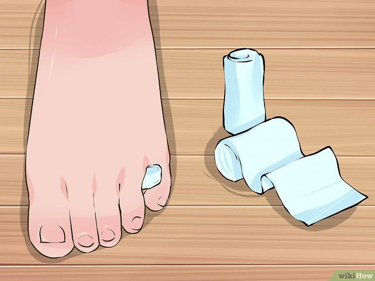 Before it arrives, a cold compress can be applied to limit movement of the injured limb.
- Rest: when applying a cold compress, the limb should not move, so as not to aggravate the injury.
- Cold compress: As a rule, an ice compress can be applied to a broken foot, securing it with the surface of the tissue and not exceeding a time of thirty minutes. Such a compress helps to reduce swelling, does not worsen the condition and does not negate the effect of analgesic medications.
Treatment
Treatment of foot fractures directly depends on the type of fracture and trauma surgery on the codetal and lateral plates, as well as the introduction of chains of the bones of the fingers. After the operation, rehabilitation treatment is required.
Rehabilitation
Restoration of mobility and removal of swelling of the foot is achieved through exercises for a while every day. Professional massage is of great importance. After the weakening of the inflammatory process, the foot can be loaded. The rehabilitation period lasts from several months to six months; it allows you to put in order the tissue, joints and muscles, as well as increase immunity and improve blood circulation.
Surgical treatment of foot fractures
Complex or severe foot fractures may require surgery. This may be necessary in cases where conservative treatment does not lead to an improvement in the patient’s condition, or in the presence of bone displacement, violation of the integrity of bone tissues and damage to blood vessels and nerves.
After the operation, it is necessary to carry out rehabilitation measures, which include physiotherapy, massage, therapeutic exercises and other methods aimed at restoring the functionality of the foot. The duration of rehabilitation depends on the nature and severity of the fracture, the patient’s age and general health.
- Foot surgery is a serious intervention and requires true professionals.

- Before surgery, it is necessary to carry out all the necessary diagnostic measures and prepare the patient for the procedure.
- After the operation, the foot should not be loaded and the medical prescriptions of the doctor should be followed in order to prevent the recurrence of the fracture or other complications.
Non-surgical treatment of foot fractures
Foot fractures can be treated both surgically and non-surgically. In some cases where the fracture is not too severe, non-surgical treatment may be a more appropriate option.
Non-surgical treatment of foot fractures may include the wearing of a cast or arch bandage. The cast will hold the foot in place, allowing it to heal and strengthen, while the arch cast will keep the foot in its natural position.
In general, non-surgical treatment of foot fractures can take a long time, but this treatment is usually safer and less invasive than surgery. More severe fractures, however, may require surgery.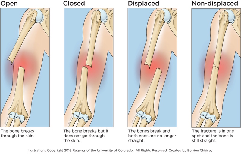
Treating the side effects of foot fractures
Weakening of the muscles and supporting ligaments
After a foot fracture, weakening of the muscles and supporting ligaments can occur, resulting in dysfunction of the foot. To restore muscle tone and strengthen the ligaments, special exercises and massage are prescribed.
Swelling and tenderness
Swelling and tenderness are natural side effects of foot fractures and can cause discomfort and difficulty in movement. To reduce swelling, drugs may be prescribed, to avoid overload. Analgesics and medications to relieve inflammation are used to reduce pain.
Gait changes and bone hypermobility
Often after a foot fracture, gait changes and bone hypermobility occur, resulting in additional pain. To solve this problem, orthopedic insoles are prescribed and physiotherapy exercises are carried out to strengthen muscles and ligaments.
Post Traumatic Stress Syndrome
People who have suffered a broken foot may develop emotional PTSD. To treat this condition, therapeutic sessions, complex breathing exercises and meditation can be prescribed.
Rehabilitation after a broken foot
Rehabilitation after a broken foot plays a key role in restoring a patient’s health and fulfilling life. In the first days after the fracture, a set of measures is prescribed to reduce swelling and pain in the area of injury. In addition, to speed up recovery, special orthopedic products are selected that provide maximum comfort and support for the injured foot.
The following stages of rehabilitation are aimed at restoring the motor functions of the foot. Physical rehabilitation specialists are involved with the patient, who select the optimal exercises and treatment methods to achieve the best results. In the process of training, the patient learns the correct walking technique and strengthens the muscles of the foot, which helps to eliminate pain and restore foot mobility.
An important component of rehabilitation after a broken foot is nutrition. The patient requires a complete diet rich in proteins, calcium and other vitamins and minerals necessary for bone healing and tissue renewal.
The rehabilitation process after a broken foot can take a long time, but a properly selected set of measures helps to return to a full life and forget about past injuries.
Possible complications of foot fractures
Infections
In case of a fracture of the bones of the foot, an infectious process may develop, which may occur as a result of injury and contact of the wound with bacteria of the internal or external environment, as well as as a result of surgical intervention.
Infectious complications can lead to an increase in body temperature, deterioration of the condition, and even threaten the patient’s life.
Neurological complications
Fracture of the bones of the foot can be accompanied by damage to the nerve structures, which can lead to impaired sensation, swelling of tissues and even paralysis of the limb. Significant nerve damage can lead to loss of limb function and disability.
Parenchymal complications
Fracture of the bones of the foot may damage internal organs such as the carotid gland and liver, which can lead to impaired function. Also, strong blows can cause damage to the kidneys, which can lead to acute kidney failure.
Thromboembolic complications
Prolonged physical inactivity and some drug treatments may be accompanied by the development of thromboembolic complications such as large vein thrombosis and pulmonary embolism, which threatens the patient’s life.
Prevention of foot fractures
To prevent foot fractures, a number of measures must be taken. One of the main factors contributing to fractures is the wrong footwear. Avoid wearing high heels and non-supportive soft shoes. Buy shoes with a comfortable fit, stiff soles, and good foot support.
In addition, it is important to keep your bones and joints healthy. Regular leg strengthening and coordination exercises will help keep your feet healthy and prevent injury. Do not get carried away with too intense workouts and do not overload your legs.
For those who work on their feet, it is important to choose the right shoes and take breaks to warm up the feet and prevent overwork. It is also worth avoiding an increased load on one specific area of the foot, for which you can use orthopedic inserts in shoes.
- Choose comfortable shoes with hard soles and good support.
- Keep your bones and joints healthy.
- Exercise regularly to strengthen your legs and maintain coordination.
- Do not overload your legs and take breaks at work.
- Use orthopedic inserts in shoes to avoid stress on a specific area of the foot.
Related videos:
Q&A:
What are the symptoms of a foot fracture?
Symptoms of a broken foot may include swelling, severe pain, impaired function of the foot, changes in the shape of the foot, and cracking, popping, or difficulty moving.
How to quickly start treatment for foot fractures?
Treatment of a foot fracture should begin immediately after the injury, and preferably within the first few hours. Therefore, it is important to seek medical help as soon as possible.
I have been prescribed a cast for a broken foot, how long do I need to wear it?
The time of wearing a cast in case of foot fracture depends on the severity of the fracture and can vary from several weeks to several months.
How is rehabilitation after a foot fracture?
Rehabilitation after a foot fracture includes restoring mobility, strengthening the muscles and joints of the foot, and treating the consequences of a fracture. It can take anywhere from a few months to a year or more, depending on the severity of the injury.
Is it necessary to have surgery for broken bones in the foot?
Some severe and complex foot fractures may require surgery. The operation is decided by the doctor after the patient undergoes special examinations.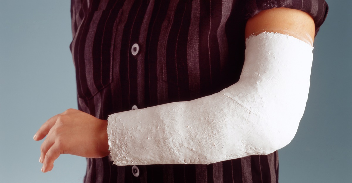
What drugs are prescribed in the treatment of foot fractures?
Foot fractures are treated with analgesics to relieve pain, anticoagulants to prevent thrombosis, drugs to speed up the healing of bones and saturate them with calcium, and immobilizers to hold the bones in the correct position, such as plaster.
Ankle malunion fracture: timely treatment prevents arthrosis
- What does an ankle malunion fracture mean?
- Complications after malunion of a fracture: Arthrosis
- What are the signs of an improperly healed fracture?
- Techniques for the treatment of malunion fractures of the ankle joint
- Which doctor performs the operation for an improperly healed fracture
- Which type of anesthesia involves surgery?
- Postoperative care, rehabilitation and aids after ankle surgery
- Will the foot hurt after the operation?
- What are the conditions for staying at the Gelenk-Klinik?
- What should I pay attention to after the surgical treatment of an ankle fracture?
- Cost of ankle fracture correction surgery
- How can a patient from abroad make an appointment for treatment of an improperly healed fracture?
X-ray of a malunion fracture. Displacement of the articular surfaces of the ankle joint. Due to the incorrect position, the load on the talus (below) is only in the region of the lateral malleolus (right). Unilateral loads can cause arthrosis. © gelenk-clinic
In case of an improperly fused fracture of the ankle joint , the bones of which it consists did not grow together properly. This leads to inevitable damage to the ankle and limitations in daily life.
The ankle joint is constantly subjected to stress. While walking through the lower leg and fibula, it takes on the full weight of the human body and transfers it to the bones of the foot. During running and jumping exercises, a force greater than body weight can be generated.
In the Gelenk Clinic you will be offered high quality treatment by the best specialists who will bring you back to your old life.
What does an malunion fracture of the ankle mean?
In the upper ankle joint, the talus is covered by the fork of the ankle. From the inside, this element forms the lower end of the tibia (tibia) and from the outside, the fibula (fibula). The upper ankle joint is responsible for moving the foot up and down. © Gelenk Clinic.
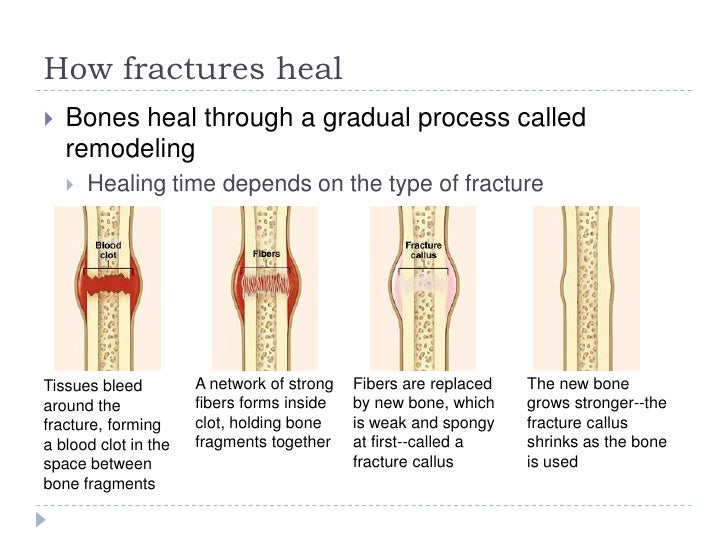
A fracture (fracture) damages the continuous structure of the bone. After a fracture, the bones can fully recover within a certain time frame. To do this, “fresh” bone material (callus) is formed between the damaged ends, which eventually becomes harder and more elastic.
The upper ankle connects the foot to the lower leg and is formed by three bones: It consists of the medial malleolus of the tibia and the lateral malleolus of the fibula. Together, these elements, like forceps, cover the wedge-shaped talus from above. A strong connective tissue apparatus leads the talus to the fork of the ankle joint.
The lower ankle joint connects the talus to the calcaneus (Calcaneus), cuboid (Os cuboideum) and navicular (Os naviculare) bones. It adjusts the position of the calcaneus and hindfoot in and out on uneven ground.
send an inquiry
Complications after an ill-healed fracture: Arthrosis
Frontal image of the foot. The lower ankle joint connects the talus to the other bones in the foot. Syndesmosis is a connective tissue connection between the tibia and fibula. It stabilizes the ankle fork. Damage to the syndesmosis leads to long-term instability. © Gelenk Clinic
Fractures of this nature can adversely affect various parts of the bones. Often, these pathologies are accompanied by injuries of the ligamentous apparatus.
If the bone fragments are not exactly parallel to each other during the healing process, deformities of the ankle joint are formed. In addition, its shape changes, which can lead to serious consequences:
- Mobility restrictions and blockades
- Pain in the foot during exertion, decline in physical activity
- Long-term ankle instability resulting in frequent twisting of the leg
- Chronic pain
- Premature wear of the articular cartilage (arthrosis) due to deformities and changes in the shape of the ankle joint
What are the types of malunion of ankle fractures?
Types of malunion fractures:
- Pseudarthrosis (false joint)
- Axial and rotational bone deformities
- Bone destruction
- Changes in bone size
- Ligament instability
Fractures of the ankle joint are fractures in the ankle region, which are divided into different types
Weber classification of ankle fractures
Fractures of the lateral malleolus are classified according to the Denis-Weber classification (often known as the Weber classification). The distribution depends on the height of the fractures in the area of the lower fibula, as well as on the damage to the syndesmosis of the ankle joint. © gelenk-clinic
- Type A: Fracture of the lateral malleolus distal to the syndesmosis (Ligamental connections between the tibia and fibula), the syndesmosis is intact.
- Type B: Fracture of the fibula at the level of the syndesmosis.
- Type C: Fracture above ankle level. There is damage to the stabilizing membrane (Interosseous membrane of the leg – lat. Membrana interossea cruris) between the tibia and fibula.
Pseudarthrosis may occur after both conservative and surgical treatment of external fractures. This term comes from the Greek (pseudo – false, arthros – joint) and describes the formation of a false joint due to improper bone healing. After a fracture, the affected fibula usually recovers slowly.
Fracture of the medial malleolus can also lead to changes in bone structures, polysegmental changes, and sometimes lengthening of the tibia.
Injuries to the posterior surface of the tibial joint (Volkmann’s triangle) can cause periostosis or dissection of the articular surface. Periostosis (layering of osteoid tissue on the cortical substance of the diaphysis of bones) leads to prolonged arthrosis of the ankle joint. Due to the accompanying ruptures of the internal ligaments, the supporting ligamentous apparatus loses its stability, and the joint is subjected to improper loading.
A special form of ankle fracture: Maisonneuve’s fracture
Maisonneuve’s fracture is an atypical, difficult-to-diagnose ankle injury. In addition to a fracture of the medial malleolus and a torn medial lateral ligament, the patient has a fibula fracture near the knee joint. Characteristic of this pathology is the rupture of the syndesmosis, as well as damage to the stabilizing membrane between the two bones of the lower leg. One of the possible complications may be deformity in the area of the “fork” of the ankle joints.
Send an inquiry
How is an ankle malunion fracture diagnosed?
The consequences of an ill-healed fracture can be seen both shortly after the injury and several decades later. Patients suffering from this pathology, as a rule, talk about noticeable ankle mobility disorders, and for some it becomes painful to step on the foot.
Six months after treatment, patients can often return to the same load on the foot, as well as play sports.
Typical symptoms of an unsuccessful ankle fracture:
- Swelling
- Foot limitation
- Blockade during flexion and extension
- Severe pain in the leg, reduced walking distance
- Prolonged instability of the joint, patients often stumble
- Chronic pain
- Limited mobility and complaints of pain in the foot lasting more than six months
- Infectious diseases in the affected area
Send request
Methods for the treatment of failed fractures
A detailed medical examination is the basis for further treatment of an improperly healed ankle fracture.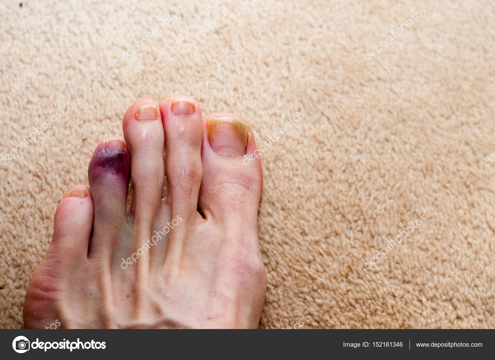 © joint-surgeon
The goal of quality treatment of patients with malunion fracture is to restore the natural functions of the ankle. How successful this is depends on the following factors:
- Degree of fractures
- Associated injuries of articular cartilage on the articular surface
- Soft tissue injuries
- Scarring
- Age and sex of patient
- Choice of conservative or surgical treatment of the ankle joint
Osteotomy: elimination of functional disorders of the ankle joint
Up to which stage of the disease a joint-sparing operation should be performed is decided by our clinic specialist on an individual basis. Together with the patient, we discuss all the details and the prognosis of joint-preserving therapy, based on existing injuries in the ankle joint.
It is worth noting that surgical treatment aimed at preserving the functions of the ankle joint by correcting its position cannot restore its natural functions in full. Even after a successful operation, the patient may experience limited mobility or sensitivity disorders in the foot.
Despite this, surgery provides marked long-term improvement in symptoms, even in patients with high emotional stress due to progressive ankle wear. In such cases, the doctors at the Gelenk Klinik eliminate the causes of arthrosis during surgery using a technique such as corrective osteotomy. Depending on the position of the fracture, the surgeon performs an operation on the outer or inner ankle or on the articular surface of the tibia.
Left: The tilt of the talus reduces the likelihood of optimal position of all components of the ankle joint. Most of the weight is on the inner (medial) edge of the talus (pointer). Due to chronic overload, arthrosis appears in this place. Right: after surgical correction of the tibia (tibia), the load on the entire articular surface is in full (pointer). Depending on the degree of development of arthrosis, osteotomy helps to stop the disease for several years. © gelenk-clinic
During this intervention, the specialist purposefully cuts the leg bone and, if necessary, removes the bone wedge. After the bone fragments are placed in the desired position and the length and position of the fibula are checked, the bone fragments are fixed using metal screws and plates.
This technique, aimed at restoring and healing the affected bones in a new position, allows specialists to restore the anatomical shape, as well as the load capacity of the ankle joint
During the operation, the surgeon diagnoses the entire ligamentous apparatus. Ruptures of syndesmosis ligaments, internal as well as external ligaments are carried out with the help of reconstructive intervention.
Autologous chondrocyte transplantation – regeneration of the articular surface
Localized injuries of the articular cartilage in an malunion of the ankle joint are eliminated by highly qualified surgeons of the Gelenk Clinic using autologous chondrocyte transplantation. This intervention involves taking cartilage cells where the articular cartilage is not damaged. In the laboratory, cartilage cells are propagated and implanted at the site of cartilage damage after about 6-8 weeks.
Ankle arthroplasty
The aim of arthroplasty is to restore painless and natural movement in the ankle joint in case of progressive arthrosis. Gait preservation is an advantage of this intervention.
Prosthesis placement requires correct position of the joint. Ligament injuries, bone changes or fractures must be eliminated in advance. This means that joint-preserving surgery (osteotomy) may precede arthroplasty. Sometimes osteotomy improves the patient’s pain symptoms and delays the need for arthroplasty surgery indefinitely.
In the absence of a recommendation for arthroplasty, the specialists of Gelenk Clinic recommend the method of immobilization (arthrodesis) of the ankle joint.
Arthrodesis – immobilization of the ankle joint
Arthrodesis is artificially created bone adhesions between the bones of the leg and foot. During an operation to immobilize the upper part of the ankle, the talus is fixed with special screws in the fork of the ankle joint going from above. After arthrodesis, the bones fuse together. Mobility in the joint is eliminated in favor of a stable bony connection. As a result of arthrodesis, the patient gets the opportunity to carry out the previous loads again without pain. Arthrodesis also eliminates the existing instability of the joint.
However, this operation is associated with significant mobility restrictions. The patient’s gait may change. Another sequence of movements leads to an overload of adjacent joints, which can cause arthrosis of the hip and ankle joints, as well as the heel region.
Send an inquiry
Which doctor specializes in performing the operation?
One of the peculiarities of the Gelenk Clinic is the relationship of trust between doctors and patients. That is why your attending physician will take care of you from the first examination to the operation itself. He will also monitor your condition after the operation. Thus, in the Gelenk-Klinik you will have a contact person to whom you can contact at any time convenient for you. The specialists of the Gelenk Clinic for the treatment of ankle malunion fractures are Dr. Thomas Schneider and Dr. Martin Rinio. These experienced surgeons are constantly improving their skills and are members of the non-profit German Association for Foot and Ankle Surgery (D.A.F). Orthopedic Clinic Gelenk Clinic in Germany is a specialist center for the treatment of foot and ankle diseases.
What type of anesthesia does surgery require?
Foot surgery is performed under general anesthesia. However, in order to avoid general anesthesia, if the patient wishes, we also offer spinal anesthesia. To do this, the anesthesiologist uses a combination of anesthetics, specially selected for the patient, and injects the anesthetic into the spinal canal of the lumbar spine. The patient is fully conscious during the operation. Together with your anesthetist, you will decide which type of anesthesia is best for you.
Send an inquiry
Postoperative care, rehabilitation and aids after ankle surgery
After ankle surgery , only partial loads are allowed. Therefore, you will need crutches. In addition, a special orthosis-fixator will be made for the patient, which he will need to wear day and night. Specialists of the Gelenk Clinic will make sure that you receive the necessary aids after the operation to correct the fracture.
Therefore, prevention of thrombosis (eg using heparin/enoxaparin) is vital. This will prevent the formation of dangerous blood clots. We strongly recommend that you undergo physical therapy and lymphatic drainage after you leave the hospital. In this way, we can prevent muscle loss and minimize swelling in the forefoot. The duration of swelling of the foot depends on the age of the patient.
Send an inquiry
Will I feel pain after the operation?
Every operation and every fracture is associated with some kind of pain – and ankle surgery is no exception. As a rule, we try to keep pain to a minimum. In most cases, the anesthetist will give a special injection that will relieve the knee for about 30 hours. After that, the pain is significantly reduced and the patient’s treatment is continued with conventional drugs. The main thing for us is to ensure a painless postoperative period.
What are the conditions for staying at the Gelenk-Klinik?
Private room at the Helek Clinic in Gundelfingen, Germany. © joint-surgeon
During your stay at the Gelenk Clinic you are usually in a private room with a shower and toilet. In addition, we provide towels, bathrobes and slippers. You can also use the safe and minibar. All rooms are equipped with a TV. You only need to take medicines, comfortable clothes and nightwear with you. Patient care is provided around the clock. The attending medical staff, as well as the physiotherapists of the Gelenk Clinic will always answer all your questions. Generally, the hospital stay after ankle surgery is 3 days. Your relatives can stay at a nearby hotel. Our staff will be happy to take care of your room reservation.
Send an inquiry
What should I pay attention to after an ankle fracture surgery?
Depending on the treatment technique, we give the following recommendations:
Osteotomy:
- Loads: Partial loads approx. 20 kg for 12 weeks
- Orthoses VACOped wear 12 weeks
- Hospital stay: 5 days
- Optimal hospital stay: 12 days
- When you can return home: 10 days after surgery
- When recommends leaving the clinic: 10 days after surgery
- When showering is allowed: 12 days after intervention
- How long is it recommended to stay on sick leave: 6-12 weeks (depending on professional activity)
- When stitches are removed: after 12 days
- When you can drive again: after 4 months
- Rehabilitation: can start after 12 weeks
- Sport: not earlier than one year later
Endoprosthetics:
- Loads: Partial loads approx.
 from 20 kg until complete healing of the wound, then a gradual increase in loads
 - Hospital stay: 5 days
- Optimal hospital stay: 12 days
- When you can return home: 10 days after surgery
- When recommends leaving the clinic: 15 days after surgery
- When you can take a shower: after 12 days
- How long is it recommended to stay on sick leave: 6 weeks (depending on professional activity)
- When stitches are removed: after 12 weeks
- When you can drive again: after 8 weeks
- Rehabilitation: can start after 8 weeks
- Sports: not earlier than one year later
- Sports: soonest after one year
Arthrodesis:
- Loads: Partial loads approx. from 20 kg for 8 weeks, then x-ray
- VACOped orthoses wear 8 weeks
- Hospital stay: 5 days
- Optimal hospital stay: 12 days
- When you can return home: 10 days after surgery
- When recommends leaving the clinic: 15 days after surgery
- When you can take a shower: after 12 days
- How long is it recommended to stay on sick leave: 8 weeks (depending on professional activity)
- When stitches are removed: after 12 weeks
- When can you drive again: after 10 weeks or depending on x-ray results
- Rehabilitation: can start after 12 weeks
- Sport: not earlier than one year later
Send an inquiry
Cost of ankle fracture correction surgery
In addition to the cost of the ankle surgery itself, additional costs for diagnostics, medical appointments and assistive devices (e. g. elbow crutches) should be taken into account, starting from about 1.500 up to 2.000 euros. If you plan to undergo outpatient physiotherapy after surgery, we will be happy to provide you with a preliminary cost estimate.
How can a patient from abroad make an appointment for treatment of an improperly healed fracture?
To begin with, the specialists of the Gelenk-Klinik will need up-to-date MRI and X-rays in order to assess the condition of the ankle joint. You will be emailed patient information as well as cost estimates.
Foreign patients can book an ankle fracture repair surgery in a short time. We will be happy to assist in obtaining a visa, after the prepayment indicated in the estimate has been credited to our account. In the event that a visa is not issued, the amount received will be returned to you in full.
Due to sometimes long flights, we try to keep the time between the first examination and the operation to a minimum. During outpatient and inpatient treatment of the ankle joint, you will be able to use the services of qualified medical personnel who speak several foreign languages (eg English, Russian, Spanish, Portuguese).

 However, they must ensure the bandage is not too tight, as this may stop the blood from circulating.
 9 Rehabilitation after a foot fracture
 Occurs with a significant impact on the foot and may be accompanied by a bone fracture.

 from 20 kg until complete healing of the wound, then a gradual increase in loads