
Lacerations: Medical Term for Gash – Symptoms, Causes, and Treatment
What are the symptoms of a laceration. How to treat a gash at home. When should you seek medical attention for a cut. What are the best ways to prevent lacerations.
Understanding Lacerations: The Medical Term for Gash
A laceration, commonly known as a gash, is a type of wound characterized by a tear or opening in the skin. This injury can vary in severity, from superficial cuts to deep wounds that affect underlying tissues. Understanding the nature of lacerations is crucial for proper treatment and prevention of complications.
Types of Lacerations
- Superficial lacerations: Affect only the outer layer of skin
- Deep lacerations: Penetrate beyond the skin into deeper tissues
- Complex lacerations: Involve damage to nerves, blood vessels, or tendons
Are all cuts considered lacerations? Not necessarily. While all lacerations are cuts, not all cuts are classified as lacerations. The term “laceration” typically refers to a more severe injury with jagged or torn edges, often caused by blunt force trauma or sharp objects.

Recognizing Laceration Symptoms and Assessing Severity
Identifying the symptoms of a laceration is crucial for determining the appropriate course of action. The severity of a laceration can range from minor to life-threatening, depending on various factors.
Common Symptoms of Lacerations
- Bleeding, which can be mild to severe
- Pain or discomfort around the wound site
- Visible tear or opening in the skin
- Redness and swelling around the affected area
- Difficulty moving the injured body part (if deep tissues are affected)
How can you assess the severity of a laceration? Consider the following factors:
- Depth of the wound
- Location on the body
- Amount of bleeding
- Presence of foreign objects
- Signs of infection
Causes and Risk Factors for Lacerations
Lacerations can occur due to various reasons, ranging from everyday accidents to more severe incidents. Understanding the common causes can help in prevention and prompt treatment.
Common Causes of Lacerations
- Accidents with sharp objects (knives, glass, tools)
- Falls or collisions resulting in skin tears
- Sports-related injuries
- Workplace accidents
- Motor vehicle accidents
Who is at higher risk for lacerations? While anyone can sustain a laceration, certain factors may increase the risk:

- Age: Both young children and older adults are more prone to injuries
- Occupation: Those working with sharp tools or machinery
- Participation in high-risk activities or sports
- Medical conditions affecting balance or coordination
First Aid and Home Treatment for Minor Lacerations
Proper first aid is crucial in promoting healing and preventing complications for minor lacerations. Here’s a step-by-step guide for treating minor cuts at home:
- Clean your hands thoroughly with soap and water
- Rinse the wound under cool running water for several minutes
- Clean the surrounding skin with mild soap
- Apply gentle pressure with a clean cloth to stop bleeding
- Apply an antibiotic ointment to prevent infection
- Cover the wound with a sterile bandage or gauze
Is it necessary to use hydrogen peroxide or alcohol to clean a laceration? Contrary to popular belief, these substances can actually harm the tissue and delay healing. Stick to plain water and mild soap for cleaning minor wounds.
When to Seek Medical Attention for Lacerations
While many minor cuts can be treated at home, certain situations require professional medical care. Recognizing these scenarios is crucial for proper wound management and prevention of complications.
Signs That Medical Attention Is Needed
- Uncontrolled bleeding after 10-15 minutes of direct pressure
- Deep wounds that may require stitches
- Wounds with visible fat, muscle, or bone
- Injuries from rusty or dirty objects
- Animal or human bites
- Signs of infection (increased pain, redness, swelling, or fever)
- Wounds on the face or near joints
How soon should you seek medical care for a serious laceration? For severe injuries or those showing signs of infection, it’s best to seek medical attention as soon as possible, ideally within a few hours of the injury occurring.
Professional Treatment Options for Lacerations
When a laceration requires medical attention, healthcare professionals have several treatment options available, depending on the severity and location of the wound.
Common Medical Treatments for Lacerations
- Wound cleaning and debridement
- Sutures (stitches) or staples
- Skin adhesives (medical glue)
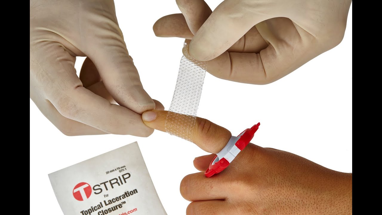
- Steri-strips or butterfly closures
- Tetanus shot, if necessary
- Antibiotics for infection prevention or treatment
How do doctors determine the best treatment for a laceration? Factors considered include the wound’s depth, location, cleanliness, and the time elapsed since the injury occurred. The patient’s overall health and risk factors also play a role in treatment decisions.

Preventing Complications: Proper Wound Care and Follow-up
Proper wound care is essential for preventing complications and promoting optimal healing. Whether treated at home or by a medical professional, following these guidelines can help ensure a smooth recovery:
- Keep the wound clean and dry
- Change dressings regularly, as directed
- Avoid picking at scabs or removing stitches prematurely
- Monitor for signs of infection
- Follow any prescribed medication regimens
- Attend follow-up appointments as scheduled
Can all lacerations heal without scarring? While proper wound care can minimize scarring, some degree of scarring is often inevitable, especially for deeper or more severe lacerations. Factors such as age, skin type, and wound location can influence the appearance of scars.
Strategies for Preventing Lacerations and Promoting Safety
Prevention is always better than cure when it comes to lacerations. Implementing safety measures and being mindful of potential hazards can significantly reduce the risk of injury.

Effective Laceration Prevention Techniques
- Use proper protective equipment when working with sharp tools
- Keep knives, scissors, and other sharp objects out of children’s reach
- Maintain a clutter-free environment to prevent trips and falls
- Exercise caution when handling glassware or fragile items
- Use appropriate safety gear during sports and recreational activities
- Ensure proper lighting in work and living spaces
How can workplaces reduce the risk of lacerations? Implementing safety protocols, providing proper training, and ensuring the use of appropriate protective equipment are key strategies for preventing workplace lacerations.
Understanding lacerations, their causes, and proper treatment methods is essential for everyone. By recognizing the signs of a serious injury and knowing when to seek medical attention, you can ensure prompt and effective care. Remember, prevention is always the best approach, so take necessary precautions to minimize the risk of lacerations in your daily life.
Lacerations, while common, should never be taken lightly. Proper assessment, treatment, and care can make a significant difference in the healing process and prevent potential complications. Whether you’re dealing with a minor cut or a more severe gash, always prioritize wound care and don’t hesitate to seek professional medical advice when needed.
By staying informed about laceration prevention and treatment, you’re better equipped to handle these injuries if they occur and to create a safer environment for yourself and those around you. Remember, a little knowledge and caution can go a long way in preventing and managing lacerations effectively.
Cuts and puncture wounds: MedlinePlus Medical Encyclopedia
A cut is a break or opening in the skin. It is also called a laceration. A cut may be deep, smooth, or jagged. It may be near the surface of the skin, or deeper. A deep cut can affect tendons, muscles, ligaments, nerves, blood vessels, or bone.
A puncture is a wound made by a pointed object such as a nail, knife, or sharp tooth. Puncture wounds often appear to be on the surface, but may extend into the deeper tissue layers.
Symptoms include:
-
Bleeding - Problems with function (movement) or feeling (numbness, tingling) below the wound site
- Pain
Infection may occur with some cuts and puncture wounds. The following are more likely to become infected:
- Bites
- Punctures
- Crush injuries
- Dirty wounds
- Wounds on the feet
- Wounds that are not promptly treated
If the wound is bleeding severely, call your local emergency number, such as 911.
Minor cuts and puncture wounds can be treated at home. Prompt first aid can help prevent infection and thereby speed healing and reduce the amount of scarring.
Take the following steps:
FOR MINOR CUTS
- Wash your hands with soap or antibacterial cleanser to prevent infection.
- Then, wash the cut thoroughly with mild soap and water.
- Use direct pressure to stop the bleeding.
- Apply antibacterial ointment and a clean bandage that will not stick to the wound.
FOR MINOR PUNCTURES
- Wash your hands with soap or antibacterial cleanser to prevent infection.
- Rinse the puncture for 5 minutes under running water. Then wash with soap.
- Look (but do not poke around) for objects inside the wound. If found, don’t remove them. Go to your emergency or urgent care center.
- If you can’t see anything inside the wound, but a piece of the object that caused the injury is missing, also seek medical attention.
- Apply antibacterial ointment and a clean bandage that will not stick to the wound.

- DO NOT assume that a minor wound is clean because you can’t see dirt or debris inside. Always wash it.
- DO NOT breathe on an open wound.
- DO NOT try to clean a major wound, especially after the bleeding is under control.
- DO NOT remove a long or deeply stuck object. Seek medical attention.
- DO NOT push or pick debris from a wound. Seek medical attention.
- DO NOT push body parts back in. Cover them with clean material until medical help arrives.
Call 911 or your local emergency number if:
- The bleeding is severe or cannot be stopped (for example, after 10 minutes of pressure).
- The person cannot feel the injured area, or it doesn’t work right.
- The person is otherwise seriously injured.
Call your health care provider right away if:
- The wound is large or deep, even if the bleeding is not severe.
- The wound is more than a quarter inch (.64 centimeter) deep, on the face, or reaching the bone. Stitches may be needed.
- The person has been bitten by a human or animal.
- A cut or puncture is caused by a fishhook or rusty object.
- You step on a nail or other similar object.
- An object or debris is stuck. Do not remove it yourself.
- The wound shows signs of infection such as warmth and redness in the area, a painful or throbbing sensation, fever, swelling, a red streak extending from the wound, or pus-like drainage.
- You have not had a tetanus shot within the last 10 years.
 Stitches may be needed.
Keep knives, scissors, sharp objects, firearms, and fragile items out of the reach of children. When children are old enough, teach them to how to use knives, scissors, and other tools safely.
Make sure you and your child are up to date on vaccinations. A tetanus vaccine is generally recommended every 10 years.
Wound – cut or puncture; Open wound; Laceration; Puncture wound
- First aid kit
- Laceration versus puncture wound
- Stitches
- Snake bite
- Minor cut – first aid
Ball JW, Dains JE, Flynn JA, Solomom BS, Stewart RW. Skin, hair, and nails. In: Ball JW, Dains JE, Flynn JA, Solomom BS, Stewart RW, eds. Seidel’s Guide to Physical Examination. 9th ed. . St Louis, MO: Elsevier; 2019:chap 9.
Lammers RL, Aldy KN. Principles of wound management. In: Roberts JR, Custalow CB, Thomsen TW, eds. Roberts and Hedges’ Clinical Procedures in Emergency Medicine and Acute Care. 7th ed. Philadelphia, PA: Elsevier; 2019:chap 34.
Simon BC, Hern HG. Wound management principles. In: Walls RM, Hockberger RS, Gausche-Hill M, eds, eds. Rosen’s Emergency Medicine: Concepts and Clinical Practice. 9th ed. Philadelphia, PA: Elsevier; 2018:chap 52.
Updated by: Jesse Borke, MD, CPE, FAAEM, FACEP, Attending Physician at Kaiser Permanente, Orange County, CA. Also reviewed by David Zieve, MD, MHA, Medical Director, Brenda Conaway, Editorial Director, and the A.D.A.M. Editorial team.
Lacerations: Definition, Diagnosis & Treatment | Portland Urgent Care
Lacerations are scary when they happen, especially if you do not know what to look for or how to handle a cut. Knowing what to look for and how to handle a laceration will help you to determine the correct form of treatment. Find out everything you need to know about lacerations to make the best decisions for you and your family.
What is a laceration wound?
A laceration wound refers to a skin wound without missing skin. Usually, lacerations are caused by sharp objects. These are one of the easiest medical conditions to diagnose and easy to treat. Lacerations form by tearing the soft body tissue, that is, the top layer or layers of skin. Furthermore, lacerations are irregular tear-like wounds often caused by blunt trauma.
Puncture wounds break more than soft tissue. Lacerations can be deep, shallow, long, short, wide, and even narrow. Minor lacerations do not usually require medical assistance as they can be treated at home with proper cleaning, ointments, and bandages. Also, minor lacerations will not bleed as much as deep lacerations.
Deeper lacerations may require stitches if they are deep, bleeding profusely, have jagged edges, or expose muscle or fat. Seek medical attention for deep lacerations, especially cuts that will not stop bleeding. Often lacerations are misused as incisions that are caused on purpose or have clearly defined edges.
What are the signs and symptoms of a laceration?
Lacerations are easy to spot as they refer to damage to the skin. As the skin has nerves, you will feel a sharp pain from a cut. Also, the cut skin will bleed and have a visible tear in the skin when the blood is out of the way. Often you will know when a laceration happens as it involves a cut or injury. The laceration victim will often scream in pain when the accident occurs, which is the first symptom of a cut.
How do you describe a laceration?
Describe a laceration as a defined tear in the tissue of the skin caused by either shearing or crushing force. Often, lacerations are a result of blunt trauma. A laceration can also be described as an incomplete separation of strong tissue elements such as blood vessels or nerves. Lacerations can be caused by both sharp or dull trauma.
What is the difference between a cut and a laceration?
Cuts and lacerations are often used interchangeably as both indicate damaged skin from a blunt or sharp object. However, a cut often refers to a mild laceration as cuts do not often require more than antibacterial ointment and a bandage. Lacerations may be deeper and require pressure to stop the bleeding and even stitches depending on the depth of the injury or exposure of other parts like bone, tendons, ligaments, or muscle.
How do you treat a wound laceration?
The first step to treat laceration is to stop the bleeding with pressure and gauze or bandage. Once the wound stops bleeding, clean the area to remove all dirt and debris. Clean by running cool water over the area and then use mild soap and water if possible. Dry with a sterile cloth.
Next, apply antibiotic ointment and cover the wound with a sterile gauze bandage and medical tape. For smaller lacerations, use a self-sticking bandage for the wound. Clean and replace bandages daily until the wound heals. For smaller cuts, you may be able to use skin closure strips. Avoid using liquid bandages for cuts without consulting a doctor first.
For deeper lacerations, go to the doctors for stitches. If you can see anything other than the first layer of skin, you also need to go to the doctor for proper treatment. If a cut measures larger than half an inch or has a large gaping wound, it probably requires stitches.
Moreover, the location of a wound may require stitches to stay shut, such as on a joint, face, near the eye, or in the genital area. Another indication that a laceration requires medical attention is a risk of infection or disease such as a rusty nail, a scratch or bite, or another potentially contaminated item. Finally, prolonged bleeding requires medical assistance.
After a few days, even if treating a minor laceration, you need to look for signs of infections or complications. Look for fever, chills, redness, swelling, white or yellow pus or drainage from the wound, or worsening pain. Do not wait to see a doctor if any of these symptoms occur; seek medical attention quickly.
Can a laceration heal without stitches?
Eventually, a laceration will heal on its own without stitches. However, stitches promote faster healing, keep the wound clean from bacteria and infection, and prevent scarring. Furthermore, stitches or staples can help to reduce blood loss and reduce future complications from the wound. Lacerations can sever toes or fingers, and these cannot heal well without stitches.
How long does it take for a laceration to heal?
Depending on the wound, it can take up to three months for the wound to fully heal. If you require stitches, the wound can heal faster in about six to eight weeks. Minor cuts and lacerations can heal in as little as two weeks, especially if the cut is very small.
The location of the wound can impact healing as well. If the injury is on your hand, foot, knee, or elbow, it may take longer to heal as the body part moves more often. Immobilizing and injury on these parts, while frustrating, can help the cut to heal faster.
How can I make my laceration heal faster?
The best way to help a laceration heal faster is to take proper care of the wound. Furthermore, if the cut requires stitches, then get stitches. Most importantly, keep the wound clean to prevent infection and covered to keep out dirt and debris. Avoid an unhealthy diet and drink more water to help provide the nutrients you need to heal properly. Try to eat food rich in vitamin C and antioxidants to help heal quickly. Lastly, avoid smoking and drugs of any sort to give yourself the best chance of healing.
How deep does a cut heal?
Lacerations heal in four stages. Stage one is stopping the bleeding, also called hemostasis. Adding pressure can help to stop blood flow as the blood clots to prevent blood loss and closes the wound by making a scab, which is stage two. At this stage, you may notice inflammation as well as that helps to heal.
Stage three involves rebuilding or growth as oxygen-rich red blood cells move to the injury and create new tissue. The last stage is maturation or strengthening, where the wound clots and heals. At this stage, you may notice itching or tightness around pink or stretched skin. From here, the body will continue to heal until the wound is gone or left with a scar. Over time, even deep cuts will heal, but stitches will help to reduce healing time.
How do you describe a deep laceration?
A deep laceration is a severe laceration. Describe a deep laceration by the size, size, shape, orientation, and margins. You could also describe a deep laceration as a gash as it implies a longer or deeper cut. Make sure to also describe if you can see bones, muscles, or other internal parts that should not be visible.
What is a severe laceration?
Severe lacerations are those that require stitches, are infected, or will not stop bleeding. Deep lacerations that expose internal parts are also severe. They may extend through more layers of tissue and cause significant pain. Do not hesitate to go to a doctor for a severe laceration. While minor cuts can be cared for at home, deep or severe cuts require medical attention.
Should I see a doctor for a laceration?
If, after applying pressure, the bleeding does not stop, then you need to see a doctor for a laceration. All severe lacerations require a doctor for treatment. Additionally, if you see signs of infection or if the laceration was caused by something that could cause infection, then seek medical attention. See a doctor also if the laceration is near the eye. Signs of shock warrant a visit to the doctors as well, including a weak pulse, clammy skin, or rapid breathing.
Signs of the wound reopening require a visit to the doctors as well. Furthermore, look for new or unexpected symptoms such as spasms, rigidity in the muscles, or near the wound. All of these symptoms may indicate complications that require professional care.
Why choose Portland Urgent Care for laceration treatment?
Portland Urgent Care works with a multitude of insurance companies to serve more customers. We also use a variety of integrated medical resources by combining both western and eastern medical healthcare which allows us to serve you the way your body needs.
We offer same-day and walk-in appointments for laceration for immediate care with the best doctors. Get a dedicated treatment plan to prevent infection and help lacerations heal quickly. From bandaging to stitches, we can do everything you need to help deal with the blood and pain to get you on the road to recovery.
Conclusion
Mild lacerations can be treated at home with antibiotic cream and a bandage. Deeper or severe lacerations that will not stop the bleed run the risk of infection or that are deep require medical attention. When in doubt, stop by Portland Urgent Care and let us look over your wound and help decide the best form of treatment to ensure a quick and safe recovery.
For more information on injuries, see our related blogs:
Common Causes & Effects of Neck Injuries
Neck Injury Treatment
Types of Back Injuries
How Do You Know If Your Back Injury Is Serious?
Types of Ankle Injuries
Common Types & Causes of Knee Injuries
How To Treat & Recover From Knee Injuries
Wrist Ligament Injuries
Wrist Injuries Causes & Treatment
Difference Between Sprains vs Strains
Peptic ulcer of the stomach and / or duodenum – City Hospital No.
 40
40
Sestroretsk, st. Borisova, 9
Online appointment
Search
Accessible environment
What is an ulcer?
Gastric and/or duodenal ulcer is a defect or erosion of its mucous membrane surrounded by an area of inflammation. Occurs as a result of an imbalance between the “protective” factors of the gastric mucosa such as the muco-epithelial barrier, microcirculation, gastric mucus, bicarbonates, hormones (gastrin, secretin, somatostatin), active regeneration, prostaglandins and “aggression” factors – pepsin, hydrochloric acid, Helicobacter pylori.
Incidence rate.
Despite the rapid development of modern medicine and pharmacology, the emergence of the latest medical equipment, a wide range of methods for examining and treating patients, gastric ulcer remains a fairly common disease.
Trusting the statistics, it can be argued that from 6 to 14% of the population in different parts of the world suffer from stomach ulcers.
In Russia, stomach ulcers can be found in about 10% of the population. Children account for 1% of the incidence.
Men aged 40-60 are most often affected. Rarely seen in teenagers.
Risk factors
- Genetic predisposition
- Helicobacter Pylori
- Smoking
- Blood type I antigens
- Medicines
- Alcohol abuse
- Irrational diet (spicy, salty, rough food)
- Violation of the evacuation of food from the stomach
- Reduced immunity
- Nervous and physical strain, frequent stress
- Meteorological influences (seasonal)
- Insufficient amount of vitamins in the body
Depending on the location of the lesion, four types of ulcers are distinguished:
- Type I – erosion occurs in the body of the stomach and at the site of its transition to the antrum.
- Type II – association of gastric ulcer with duodenal ulcer.
- III type – lesion of the pyloric part of the stomach.
- Type IV – ulcers that occur on the lesser curvature in the upper part of the stomach, in the area of the transition of the esophagus to the stomach. These ulcers are highly prone to malignancy.

What symptoms characterize a stomach ulcer?
Pain in the abdomen (in the epigastric region). By nature, these pains are burning, aching, pressing, squeezing. The pain radiates to the region of the left hypochondrium, lower back on the sides of the spine. Duration from 90 minutes to 3 hours. Characterized by seasonal exacerbation of pain (spring, autumn).
- Nausea
- Heartburn
- Belching (air or food)
- Vomiting (very rare)
- Constipation
- Weight loss
- Disorder of appetite (more often it increases)
Due to unbearable pain in the epigastric region, patients have to take a forced position: squatting, they grab their stomach with both hands or press against the edge of the table, lying in bed, turn on their stomach, etc.
What can be visually detected in a patient with a stomach ulcer?
- White coated tongue
- Excessive sweating, wet palms
- Anterior abdominal wall very sensitive
- Sharp pains on pressure in the epigastric region
- Appearance of tender points on the back in the region of the spine
How to diagnose peptic ulcer?:
- Complete blood count (remains unchanged when the disease is not complicated by other diseases)0037
- Fecal occult blood test – Gregersen test
- Study of the acid-forming function of the stomach (intragastric pH-metry)
- Detection of Helicobacter pylori
- X-ray method: using a contrast agent, this method allows you to detect defects in the gastric mucosa.
FEGDS (fibroesophagogastroduodenoscopy) with a biopsy from the bottom of the ulcer from 4-6 points and a mandatory cytological examination of the biopsy.
Ultrasonography (based on the use of ultrasound at a frequency of approximately 30,000 Hz to image deep body structures) identifies a defect in the stomach wall.
Electrogastroenterography (a method designed to study the motor-evacuation function of the gastrointestinal tract, based on the simultaneous registration of biopotentials from different parts of the gastrointestinal tract).
Possible complications
In most cases, an early diagnosed gastric ulcer can be cured without the development of any complications. But in those cases when the patient begins to neglect his health (the unwillingness of the patient to see a doctor, relying on the fact that the pain will pass by itself; fear of the upcoming diagnosis and procedures; material problems, etc.), he makes his body suffer more and more , which leads to the development of terrible complications:
- Bleeding
- Penetration (penetration of an ulcer into surrounding tissues and organs)
- Perforation (perforation of the ulcer)
- Peritonitis (spread of infection in the abdominal cavity, inflammation of the peritoneum)
- Perivisceritis (formation of adhesions to neighboring organs)
- Ulcer malignancy (malignancy)
Conservative treatment
- Substances that neutralize hydrochloric acid.
- Drugs that suppress the secretion of gastric juice inhibitors of the proton pump.
- Helicobacter pylori preparations
- Drugs that stimulate “protective” factors.
- Sedatives.
- Antispasmodics.

Surgical treatment
Subtotal resection of the stomach, gastrectomy, vagotomy. Apply only according to indications (bleeding from the upper gastrointestinal tract, perforation, penetration, stenosis of the pyloric sphincter, ulcer of endocrine origin – gastrinoma).
CHI
VHI
Paid
Desired visit date
your name
Your last name
Your patronymic
your phone number
A comment
I agree to the processing of personal information
Make an appointment
Appointment for a consultation by phone +7 (812) 200-16-88
Department of paid services: +7 (812) 437-11-00 and +7 (911) 766-97-70
Clinical Hospital | Acute stomach and duodenal ulcers
What is peptic ulcer of the stomach and duodenum?
An ulcer (ulcer in Latin) is a defect in the stomach wall that penetrates deeper than the mucous membrane. Sometimes an ulcerative defect extends through all layers of the stomach into the abdominal cavity (such an ulcer is called perforated or perforated).
In rare cases, an ulcer can penetrate into other organs, forming an inflammatory infiltrate, penetrating the intestines, omentum, liver, pancreas. This complication of peptic ulcer disease is called penetration.
Superficial defects of the gastric mucosa are called erosion. Unlike ulcers, erosions are much less likely to lead patients to severe consequences in the form of bleeding or perforation. However, they also require attention and treatment from a specialist doctor.
What are ulcers?
There are ulcers acute i.e. formed in a short period of time, sometimes in a few hours, and chronic that exist for months and years. If the ulcer does not heal within 2 to 3 weeks of starting drug therapy, it becomes chronic.
Acute ulcers are much more likely to lead to a sharp deterioration in a person’s condition. This is manifested by sharp pains in the upper abdomen, the stomach becomes overly tense and painful, surgeons call such a stomach “board-shaped”. Sometimes there is increasing weakness, nausea and vomiting with black masses of the “coffee grounds” type, black tarry stools – these signs speak in the resulting ulcer bleeding. Without the provision of emergency surgical care, such conditions lead to severe complications and death, even in modern reality.
A long-term ulcer is a chronic seasonal relapsing disease of the stomach and duodenum, which is characterized by the absence of complete healing of the ulcer or its recurrence after healing (relapse).
Peptic ulcer is characterized by periods of exacerbation (spring and autumn) and remission, when the symptoms subside.
Why is the disease dangerous?
Peptic ulcer is complicated by bleeding from an open peptic ulcer, formation of a through hole (perforation) in the stomach wall with spillage of stomach contents and hydrochloric acid into the abdominal cavity, cicatricial narrowing of the duodenal lumen. If an ulcer exists for more than 10 years, the risks of transforming the ulcer into a malignant tumor of the stomach or duodenum increase significantly.
Surgeons and endoscopists of the Federal State Budgetary Institution “Clinical Hospital” have extensive experience in the treatment of peptic ulcer and provide highly qualified medical care around the clock.
What are the symptoms of gastric ulcer and duodenal ulcer?
- Aching or cramping pains, of low intensity in the epigastric (pit of the stomach), more often occur on an empty stomach or immediately after eating
- Persistent heartburn, especially at night and in the morning
- Loss of appetite
- Nausea, sometimes vomiting
- Belching of rotten air, sour or bitter taste
- Heaviness in the epigastric region after eating, feeling of rapid filling of the stomach
- Bleeding from ulcers results in coffee grounds vomiting, black tarry stools (melena), increasing general weakness, malaise
- With perforation of ulcers – severe, dagger pain in the epigastric region, nausea, vomiting, painful tension of the abdominal muscles, a sharp deterioration in the general condition.
Causes of peptic ulcer disease
- Presence of resistant Helicobacter pylori bacteria in the stomach and duodenum, which is the main etiological factor in the occurrence of ulcers
- Violation of the diet
- Alcohol and tobacco abuse
- Long-term use of drugs without medical supervision
- Emotional and physical overstrain
- Genetic predisposition
- Metabolic disorders
- Hypovitaminosis.
Diagnostics
The main method for diagnosing gastric and duodenal ulcers is endoscopic fibrogastroduodenoscopy (EGD). The examination is carried out using a flexible endoscope with a diameter of 5 to 10 mm, which is carried out under conditions of light anesthesia, through the patient’s mouth into the esophagus, and then into the stomach and duodenum. The endoscope camera captures changes in the mucous membranes and broadcasts them on the screen. If necessary, during the study, the doctor can take a piece of tissue from the ulcer for histological examination, perform an endoscopic stop of bleeding by chipping a bleeding vessel in the ulcer or electrocoagulation. In some cases, a special mini-clip is applied to the vessel.
Necessary types of additional examinations for peptic ulcer of the stomach and duodenum are:
- General analysis of blood and urine
- Biochemical blood test
- Coagulogram
- Fecal occult blood test
- ECG
- Abdominal ultrasound
- X-ray of chest and abdomen
- General practitioner, gastroenterologist consultation
Treatment of peptic ulcer of the stomach and duodenum
Methods for the treatment of peptic ulcer are divided into conservative and surgical. In the case of an uncomplicated ulcer for the first time, treatment is carried out by prescribing drug therapy along with a mandatory diet. An important point is the exclusion of serious personal stress in the patient.
Currently, so-called multicomponent treatment regimens (three, less often four-component) are used.
In the treatment of peptic ulcer, the following drugs are prescribed:
- Antisecretory drugs (help suppress the secretion of hydrochloric acid): proton pump inhibitors (omeprazole, pantoprazole, esomeprazole, etc.)
- Antacids (Phospholugel, Almagel, etc.)
- Antibacterials, if Helicobacter
- Prokinetics (contribute to the acceleration of the movement of the food bolus from the esophagus to the stomach, then to the duodenum, increase the tone of the lower esophageal sphincter): domperidone (motilac, motilium), itopride (ganaton)
is detected
The treatment regimen is selected individually by a gastroenterologist in accordance with the patient’s condition and concomitant diseases.
Surgical treatment of peptic ulcer
Surgical treatment of peptic ulcer is required in case of complicated course of the disease. Most often, this need arises when a patient is suspected of perforating an ulcer with the development of peritonitis and ulcerative bleeding. In such cases, only emergency surgical care helps to avoid serious complications.
The patient is urgently undergoing endoscopic fibrogastroduodenoscopy under general anesthesia, in which an ulcer is detected and the type of complication is determined, as well as the amount of assistance needed. If acute ulcerative bleeding is detected, endoscopic hemostasis is performed by chipping the ulcer with vasoconstrictor drugs, or bleeding is stopped by argon-plasma coagulation, after achieving satisfactory hemostasis in a stable condition, the patient is transported to the ward for dynamic observation. In the future, the patient’s general condition is monitored, laboratory control, fibrogastroscopy control, approximate hospitalization time is 7-8 days, depending on the severity of the ulcer.
If it is impossible to stop bleeding by endoscopic method, an emergency operation is performed, the main task of which is to suture the bleeding vessel or resect part of the stomach.
If a perforated ulcer is suspected, the patient undergoes an emergency x-ray examination of the abdomen, which in most cases indicates the presence of free gas in the abdominal cavity. This sign indicates a defect in the wall of a hollow organ and requires emergency surgery. In some cases, it is necessary to perform computed tomography as a more effective method for diagnosing complex cases.
Perforation of the ulcer causes chemical and then bacterial peritonitis, which, if not treated in time, can lead to death! The time interval from the onset of the disease to the operation is very important here! Severe pain syndrome quickly exhausts the patient, and the growing general deterioration of the condition further worsens the situation.
During perforation, the pain syndrome is pronounced, the patient has a dagger pain in the abdomen, the patient cannot find a place for himself, cannot stand up – “Vanka-Vstanka symptom”. The only cure for this complication of peptic ulcer is emergency surgery.
A diagnostic laparoscopy is performed on the patient in the operating room under general anesthesia. Through several punctures of the anterior abdominal wall, air or carbon dioxide is injected into the abdominal cavity. Then a mini-camera and special 5 mm instruments are inserted. After the initial examination of the abdominal cavity, a decision is made on the implementation of one or another type of surgical assistance. If there is a through hole in the duodenum or stomach, it is sutured and the entire abdominal cavity is washed with special solutions to clean water. In parallel, a special thin probe is inserted into the patient’s stomach to protect the area of the sutured defect and drain gastric juice. The probe can serve as a way for the introduction of food after surgery and is usually removed on the first or second day. The operation ends with the installation of one or more silicone drainage tubes. This ensures the outflow of inflammatory contents from the abdominal cavity for several days.
In some difficult cases, in the presence of generalized long-term peritonitis or technical difficulties, surgeons resort to open surgery by performing a median incision in the abdominal wall.
Approximate terms of hospitalization of the patient during laparoscopy are 4-7 days. In the case of open intervention, they depend on the severity of the patient’s condition.
With long-term ulcers of the stomach and duodenum, narrowing of the lumen of these organs may form, making it difficult for food to pass through them. This condition is called stenosis. Stenosis may be complete or incomplete. With complete stenosis, the patient cannot feed on his own. Eaten food partially or completely comes out with vomit, the patient loses weight, loses strength and desire to work, live.
This situation requires urgent hospitalization and medical support and special medical nutrition (nutrition therapy). The patient needs to quickly establish a full intake of proteins, fats and carbohydrates by installing a nutritious gastric tube and intravenous nutrition.